
Visualising nature: Models and wall charts for teaching biology in Australia and New Zealand
Introduction
In the late 19th century, biology emerged as a professional scientific discipline anchored in comparative anatomy and morphology, the study of form and its development. With the impetus of Darwinian evolutionary ideas and improved histological techniques, the investigation of internal structures and evolutionary embryological development brought a new scientific biology, one which moved away from descriptive classification. This new biology took place primarily in the university laboratory, led by innovations in German universities, which, as Lynn Nyhart has observed, ‘provided the model of the “research imperative” that would be imitated by others around the world’.[1] Natural history, its predominance usurped by academic biology, continued largely outside universities, mainly located in large public museums focused on systematics research based on their specimen collections.
Teaching biology in the university laboratory required more than collections of specimens and books; it required a new type of scientific illustration. Modern biology demanded sophisticated observational capabilities, grounded in skilled microscopy. It was an area of complex technical detail: scientists were interested in internal structures and development, which often could not be seen with the naked eye. Students had to be taught what to see through the microscope. Learning through the eye became the path to understanding. Into classrooms once dominated by dry verbalism, scientific visual aids were introduced as part of instruction by seeing and doing, building upon the pedagogical theories of Swiss theorists Johann Heinrich Pestalozzi and Philipp Emanuel von Fellenberg developed in the early 19th century, which promoted education by observation.[2] This new method of teaching based on the student as the active investigator was introduced into the new biological laboratories.
The use of educational models and wall charts to teach animal and plant anatomy and morphology was not, however, solely within the purview of the universities and colleges. Museums, a number of which operated in close connection with the universities, increasingly took on an educational role for the public, employing scientific visual aids as a key form of instruction, while contributing to their own taxonomic- based collection research. By the end of the 19th century, such visual aids, as Tony Bennett has noted, were ‘widely considered crucial to the pedagogic role of a museum’ inspired by the modern systems of display initiated at the South Kensington Museum, further underscoring the belief that learning and understanding could be done through the eye.[3] Algernon Thomas, the chair in Natural Science at Auckland University, in an address to the Australian Association for the Advancement of Science in Melbourne in 1890, stressed the importance of ‘a properly displayed and explained museum in advancing biological science’[4] and in making it comprehensible to the general public.
In the lecture hall and laboratory, models and wall charts sat with lantern slides, the blackboard, and specimens as tools for teaching biology. While employed at a variety of educational levels, these visual aids were vehicles for scientific ideas, and were often produced in collaboration with scientists who worked closely with manufacturers and illustrators to give visual expression to their ideas. Models and wall charts took on the significant role of visualising the fugitive organic matter of soft-bodied invertebrates, plants, and microscopic organisms that were difficult to preserve with their texture, colour, and shape intact.[5] Their scientific accuracy and fidelity to nature were their key selling points; they were also governed, however, by a strong artistic component inspired in part by Alexander von Humboldt’s marriage of empirical observation with aesthetic appreciation.[6] They were judged on their distinct aesthetic attributes in addition to their scientific authority, which contributed to their efficacy as teaching aids.
Models were magnified into standardised forms that materialised what was difficult to understand in two dimensions, while wall charts provided an overview of external and internal structures that could be viewed from every corner of the lecture hall.[7] More specifically, models were valued for their exaggeration of detail rendered in solid three-dimensional form, which ranged from high relief to examples produced fully in the round, with the evocation of texture – through various mediums including, but not limited, to wax, wood and glass – playing a key role in the articulation of organic matter. As Nick Hopwood has observed, ‘The models’ three-dimensionality worked together with scale, texture and colour to convert delicate and shimmering but tiny and elusive forms into solid and opaque but huge and memorable shapes’.[8]
Due to the versatility of line and softness of form that characterised the lithographic medium, the aesthetic significance of wall charts lay in their ability to communicate highly legible two-dimensional illustrations of species’ gross morphological features, cellular levels and life cycles. Employed in tandem with specimens, the aim of such wall charts, as Anne Secord has demonstrated, ‘was not to teach beginners how to look at pictures but rather, how to use pictures to develop the observational skills necessary for looking at plants and other objects of nature’.[9]
The relationship between such three-dimensional and two-dimensional visual aids was complementary, forming a composite educational display that responded to contemporary print culture. Both relied for inspiration on published scientific illustrations, with drawing representing a crucial part of the transformation process from two to three dimensions. The use of colour was an important means of conveying information and was employed schematically in addition to naturally in models and charts, with colour codes often provided in publishers’ prospectuses intended for instructors.[10] While the publication of wall charts was indebted to developments in lithographic printing, models were also conceived of as a form of publication and were marketed as ‘plastic’ texts that translated living nature into visual knowledge.[11]
This process of translation through fabrication subscribed to the 19th-century cult of the copy, deriving from a long tradition of mimesis and illusion in the arts. Exactingly replicated to form perfect ‘fac-similes of nature’, scientific reproductions in the forms of models and charts were valued for their information content, truth to nature, and skill of execution over their originality.[12] While developments in lithographic printing facilitated the mechanical mass-production of large colour wall charts at reasonable prices, series of expensive models were produced or finished by hand, championing the handmade in the era of the machine-made.[13] Transforming the natural realm into a controlled and domesticated artificial kingdom, such scientific simulacra represented the triumph of form over matter and industry over nature.[14]
Within this highly industrial arena, nature flooded the interior in myriad stylised forms with scientific visual aids extending this internal migration into the laboratory and lecture hall. Models and wall charts further succumbed to current dictates of fashion and taste, and their production both contributed and responded to the rich cross-fertilisation of the intersecting spheres of science, art and design in the latter half of the 19th century, culminating in movements such as Art Nouveau or Jugendstil.
Most 19th-century models and wall charts were made in Germany, reflecting the dominance of German biologists, and were exported around the world through what Geoffrey Swinney has described as circuits of ‘production and communication’ and ‘commerce and consumption’ that revolved around mobile networks of scientists, curators, educators, illustrators, artists, craftspeople, marketing agents, shipping agents, and heterogeneous audiences who engaged with models and wall charts on multiple levels.[15] Through a preliminary analysis of European models and wall charts in Australian and New Zealand collections, this paper attempts to chart the colonial trajectory of such teaching aids and their employment in an Australasian higher educational context in universities and museums. It also seeks to uncover the rich and largely untapped local history of model and wall chart production.
While imported biological models and wall charts in colonial collections were imbued with scientific authority through their explicit connection to developing research in European university laboratories, locally produced teaching aids in Australia and New Zealand were employed to actively document, display, and promote colonial economic and agricultural development. In contrast to the male-dominated arena of production in Europe, women artists and amateurs played a more visible role in the colonial production of models and wall charts.
Models: the makers
A small number of firms dominated the biological model market, with makers engaging in direct sales, exhibiting at international exhibitions and selling through catalogues and dealers. In 1877 Ray Lankester, professor of biology at University College London, published in Nature a list of dealers he had found useful, for other scientists ‘charged with the formation of an educational museum of zoology and comparative anatomy’.[16] The leading German makers of models were featured in his list: wax embryology models by the Ziegler studio, glass invertebrate models by Leopold and Rudolf Blaschka, and wax invertebrates, especially parasitic worms, by Rudolf Weisker.[17] A significant display of German models and charts were displayed at the Exhibition of the Loan Collection of Scientific Apparatus, held at South Kensington Museum from May to December 1876, which arose out of a concern regarding the lack of science education in Britain.[18] A massive catalogue was published to accompany the exhibition, and an office set up to provide catalogues of contemporary German material with prices.
One of the most widely purchased teaching aids was the series of embryological wax models manufactured by Adolf Ziegler and his son Friedrich. Supporters of Darwinian theory saw embryology as key to proving evolutionary development, research led by German zoologists. As embryos of different species are similar in their earliest stages and are difficult to distinguish, models became a vital aid to the microscope in teaching students embryology. Based in Freiburg, the Zieglers worked with German embryologists to develop series of models of different organisms, including marine invertebrates, the frog, the chick, the beetle, and human embryos, selling their models internationally from the 1860s.
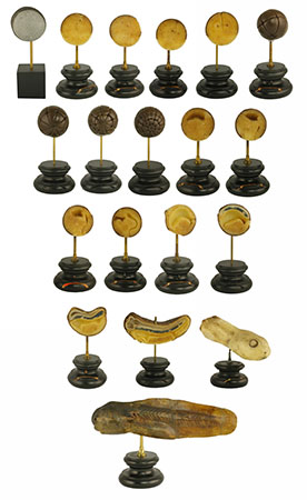
photograph by Tim Harland
Macleay Museum SC2001.36
While worked in wax, the Ziegler models were often initially conceived as drawings and continued to share an intimate relationship with two-dimensional illustrations. By the 1880s, textbooks and wall charts devoted to embryology took to reproducing figures from models, especially those made by the Zieglers, rather than plates or prepared specimens, encouraging students to compare such printed illustrations with the models themselves.[19] Instrumental not only for teaching but also for research, the Ziegler models enabled scientists to closely examine the magnified three-dimensional impressions of embryos and their substructures and even to further dissect them.[20]
The models were described in the 1876 Loan Exhibition catalogue at the South Kensington Museum as ‘scientific wax preparations for illustrating biological facts, and particularly the changes during development’ that were ‘copied from nature, and enlarged so as to be useful for teaching purposes’.[21] Emphasising the ubiquity of these German wax models, Nick Hopwood reveals that, by the turn of the century, the Zieglers supplied all German universities as well as 95 foreign universities, disseminating embryology ‘from its German powerhouse around the world’.[22]
Ziegler models were purchased between the 1880s and the early 20th century by the University of Sydney, the Australian Museum, and the University of Otago, among others. The Otago Daily Times reported in 1882 that the Otago Museum, under the curatorship of T Jeffery Parker, had acquired a collection of wax models by the Ziegler studio. In 1893 Parker exhibited a further collection of Ziegler models, ‘recently added to the museum’, at a meeting of the Otago Institute, including sets that demonstrated ‘the development of the common fowl’, the electric ray, water beetle and starfish, as well as ‘models giving greatly enlarged views of the larvae of various marine animals, and a series representing eight specimens of brains of vertebrate animals’.[23] Late 19th-century photographs of the laboratory at the Auckland University as well as references in the colonial press to the laboratory at Canterbury College suggest that Ziegler models were also present in these early collections.[24]
photograph by an Auckland Star staff photographer
Alexander Turnbull Library, Wellington, New Zealand Negatives. Ref: 1/1-002883-G
While wax was a medium conducive to embryological models, glass better suited the modelling of marine life through its ability to capture the translucence and subtle coloration of specimens. The leading glass modellers were Leopold and Rudolf Blaschka. The Blaschkas are perhaps best known for the 4000 glass models of plants, known as The Glass Flowers, commissioned by Harvard professor George Lincoln Goodale. Their initial scientific modelling business, however, was of glass marine invertebrates, specimens of which were difficult to preserve in spirits and display with their colour intact. Based near Dresden from 1863, Leopold Blaschka began with a series drawing on scientific illustrations from Philip Henry Gosse’s Actinologia Britannica: A History of the British Sea Anemones and Corals (1860), which were initially promoted as ‘decorations for elegant rooms’, subscribing to the aesthetics of the aquarium.[25] Leopold was joined by his son Rudolf in 1876 and, from 1877, the father-and-son team were in contact with German scientist Ernst Haeckel, using several of his illustrations to create sea anemones, medusa, jellyfish and radiolarians. Stylised and symmetrical, these models reveal the influence of Haeckel’s illustrative concept of Grundform, or the foundational geometric form of the organism, and Haeckel was one of the Blaschkas’ earliest customers.[26] By the mid-1880s the Blaschkas’ catalogues advertised 700 models, which were sold mostly to universities and museums both in Germany and overseas.[27]
More so than any other of the German models, their artisanal quality captured the imagination of the colonial press. An article from 1883 announces the new addition of a series of Blaschka marine invertebrate models to the Canterbury Museum in Christchurch, focusing on their technical virtuosity, stating that ‘the specimens have been placed in the Technological room in order to show them as examples of industrial art as applied to science’.[28] This consignment included corals in their natural size and in an enlarged state, the stages of growth of the jellyfish and the Physophora magnifica, and accurate depictions of the Portuguese man-of-war, the sea cucumber, eight specimens of cuttlefish, marine snails, and even the garden snail, ‘the slimy thing’ which seemed ‘about to move, it is so natural in appearance’.[29] The Australian Museum also purchased Blaschka glass marine invertebrate models in the early 1880s, a purchase explored in further detail below. In 1885 the trustees of the Queensland Museum suggested at their monthly meeting that ‘it would be expedient to procure a few of Blaschka’s well-known glass models of the lowest animals’ for ‘the student of animal life’ as they ‘render intelligible to the ordinary observer forms which cannot otherwise be presented bodily even to the naked eye’.[30]

Image from the Biodiversity Heritage Library. Digitised by WML WH01 Library
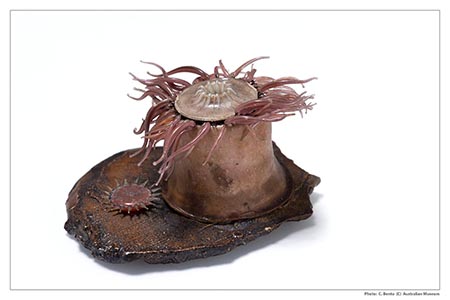
photograph by Carl Bento
Australian Museum Archives AMS582, MA817
While the Blaschkas were the leading model makers of marine invertebrate zoology to enter colonial collections, the plant kingdom was largely represented through the papier-mâchémodels of the Brendels. Robert Brendel founded his company specialising in botanical models in 1866 in Breslau, Poland, where his son Reinhold succeeded and continued the business, later moving it to near Berlin. According to the 1876 Catalogue of the Special Loan Exhibition, their models of plants, trees and flowers were used to illustrate lectures ‘regardless of the seasons, and in systematic order’.[31] Moreover, their enlarged scale was meant to ‘facilitate the recognition of all the fine and even smallest organs, and the comprehension of the distinguishing characteristics of the structure of flowers by comparison with living plants’.[32]
Made from papier-mâché and augmented with a range of other materials including feathers, cotton shirting, wood dowel, rattan, pulp cane, glass beads, gelatine and wire, the botanical models were more durable than the Blaschka models yet no less finely made.[33] Again, scientific accuracy was key. The first models were created under the guidance of biologist Ferdinand Cohn with 30 species offered for sale in 1866. The Brendels received specialist subject help from several scientists including Leopold Kny regarding the most important types of flower positions and from Carl Müller on the original forms of ovules.[34] The success of the business can be seen in that, by 1913, almost 300 species were listed and distributed widely through agents. The McGregor Museum, University of Auckland, has a splendid collection of more than 60 Brendel models, purchased in the 1920s, and a handful survive at the University of Sydney and at Otago.[35]
While most of the models came from Germany, one model maker who widely sold from France was Dr Louis Auzoux. Known for his human anatomical models, Auzoux also developed a range of papier-mâché zoological and botanical models from the 1860s. Twenty-three plant species and 24 varieties of life in large-scale models were offered for sale.[36] Models were purchased directly from the Auzoux workshop by Joseph Maiden, curator of the Technological, Sanitary and Industrial Museum (hereafter Technological Museum) in Sydney in 1883 for displays of economic botany and economic zoology.[37]
Institutions of course acquired models from multiple sources. The University of Melbourne Herbarium has 132 models made by the Brendels and Auzoux, and by Les Fils d’Émile Deyrolle in Paris.[38] The Herbarium and Botanical Museum in the Sydney Botanical Gardens, opened in 1901, held large models ‘by eminent German and French botanical model-makers’, which catered to its focus on scientific botany.[39] Teachers in applied plant sciences saw the usefulness of models of a grain of wheat and the pea pod, and insects such as the silkworm moth and honey bee, while botanical models of plants of medicinal value were desired for teaching in addition to those of agricultural use.
Models of fungi were well known to those who visited the British Museum, which purchased James Sowerby’s collection of 193 models in 1844 as well as 60 German-made models in 1888.[40] Learning the identification of poisonous and edible fungi made the models a useful tool, especially for the public. The Museum of Economic Botany in the Adelaide Botanic Gardens between 1872 and 1890 purchased over 400 fungi models from the German firm Heinrich Arnoldi & Co., then, after 1890, from Marcus Sommer.[41] Sommer’s firm, SOMSO Modelle, founded in 1876, acquired the Ziegler studio in 1936 and continues to make models under the slogan ‘nature is our model’. Auzoux also produced models of mushrooms sold through Émile Deyrolle of Paris, and possibly the Hamburg dealer Chrétien Vetter, which were acquired by the Technological Museum in Sydney.
In addition to producing botanical models, Auzoux and Sommer specialised in anatomical models of larger animals for veterinary studies, with Auzoux famously producing large papier-mâché models of horses. For the new Veterinary School at the University of Sydney, established under James D Stewart in 1910, ‘a splendid collection of skeletons and veterinary anatomical models [was] imported’, including models of parts of the anatomy of horses, pigs and sheep, examples of which still survive although their maker remains to be identified.[42]
Wall charts: the makers
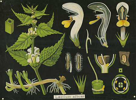
A number of the German scientists who contributed to model making also engaged in the production of educational wall charts that made their way into colonial collections. Botanist Leopold Kny, who advised the Brendels in their fabrication of papier-mâché models, also produced a series of 120 botanical wall charts on anatomy, morphology and systematics, known as Botanische Wandtafeln, published between 1874 and 1911 by the Paul Parey (and later Wiegandt and Hempel) publishing house in Berlin. Kny developed the concept of using large wall charts, measuring 69 x 85 to 106 x 150 cm, of chromolithographs mounted on linen mostly in a horizontal format for use in classroom lecture halls, which were rolled up for storage after use.[43] One of the most widely disseminated series of German wall charts, the Botanische Wandtafeln feature in the chart collection of the McGregor Museum in Auckland.
This teaching collection, which dates to 1884, also includes a series of charts on systematics by Swiss botanist Arnold Dodel-Port and his wife, Carolina, entitled the Anatomical-Physiological Atlas of Botany. Composed of 42 charts, the Atlas was designed by the Dodel-Ports who consulted a committee of botanists during the 50th Meeting of German Scientists and Physicians in Munich in 1877 regarding the selection of images.[44] In the preface to this work, the Dodel-Ports avowed, ‘Natural, scientifically reliable wallcharts can replace a natural object in classroom teaching and in lectures; they are more enlightening than the spoken word’.[45]
Other series of wall charts featuring in colonial collections include the New Wallcharts for Teaching Natural History published in 1894 and produced by teacher Heinrich Jung, college director Dr Friedrich Quentell and painter Gottlieb von Koch, who had worked as an assistant for Haeckel in Jena, before being appointed professor of zoology in Darmstadt. These are at the University of Otago, which has a handwritten catalogue of diagrams in the biological department of the university compiled in the early 20th century by William Blaxland Benham, then professor of biology.[46] Displayed on a black ground without any captions, this series of charts are not instantly intelligible, with the figures requiring verbal commentary.[47] This method made Wandtafeln incredibly versatile and adaptable yet required greater participation on both the part of the teacher and of the students.[48]
published by Frommann & M Morian, Darmstadt, Germany
Hocken Collections, Uare Taoka o Hakena, University of Otago
As with the models, descriptions were given by the teachers with the aid of explanatory booklets provided by the publishers to accompany the charts. The chart from this series on the mushroom (Agaricus campestris), in the McGregor Museum, for instance, displays handwritten annotations, particularly evident in the inscription of ‘annulus’ with an arrow pointing to the ring-like structure on the stipe (stalk) of the mushroom, suggesting that lecturers add their own labels to make the charts more readily comprehensible for students.
Lesser known botanical wall chart series linked to colonial collections include the Tabulae Botanicae, published by Gebrüder Borntraeger in Berlin, represented in the McGregor Museum, and Albert Peter’s Botanische Wandtafeln on the systematics of gymno- and angiosperms published in Berlin by Paul Parey from 1892 to 1914, which was acquired at the turn of the century by the University of Sydney. A biology requisitions book, most likely maintained by William Haswell, professor of biology, has a brief entry in 1902 recording the purchase.[49]
In addition to botanical wall charts, the McGregor Museum holds a number of late 19th-century zoological wall charts by Rudolf Leuckart, a zoologist appointed professor at the University of Leipzig in 1869, who specialised in North Sea invertebrates and in vertebrate parasitology.[50] Working with zoologist Heinrich Nitsche, Leuckart developed the Zoologische Wandtafeln, consisting of more than 100 images edited between 1877 and 1892. Like the botanical charts, they are not labelled with explanatory text, but were sold along with pamphlets intended for teachers, published in three languages: German, English and French.[51] While the styles of the charts in this series vary slightly, like the Blaschka models they are indebted to the illustrations of Haeckel, which transformed the presentation of zoology in the late 19th century, visualising the logic of nature in a form of tantalising display.[52] Their production was technically difficult and labour-intensive as they had to be printed in four sections and then joined together and touched up to achieve the large size of 1 metre by 1.4 metres.
We have canvassed here the main model and wall chart makers, whose remnant collections are found scattered in various Australasian universities, museums, and colleges. This is an area ripe for further analysis, with numerous models and charts existing in collections for which the maker is unknown or unidentified, or where some still bear original maker’s labels. Information regarding the acquisition of these teaching aids is typically scant as ephemeral purchase orders and catalogues have not often survived and, when they do, details of items procured are not outlined in payment receipts. However, surviving correspondence surrounding purchases made by the Australian Museum and the Technological Museum in Sydney through the dealers Václav Friĉ of Prague and Chrétien Vetter of Hamburg provide valuable insight into how such model and charts entered colonial collections.
Colonial acquisition of European scientific teaching aids
By the end of the 19th century, specialist science disciplines were in place in Australian and New Zealand universities. Chairs in biology had replaced chairs in natural history, and new teaching positions were filled by graduates recruited from England and Scotland who were trained in laboratory-based teaching and supported Darwin’s theory of evolution. These men were ‘thoroughly equipped to participate in the exciting and fashionable fields of comparative embryology, anatomy and physiology’ and were instrumental in shaping and transforming the study of biology in the colonies.[53]
The staff at university science departments and museums maintained close contact with their European counterparts through a network of research and personal connections that linked colonial and metropolitan centres. William Haswell, at the University of Sydney, was taught by Thomas Henry Huxley, and wrote a foundation biology textbook with Thomas Jeffery Parker, who was curator of the Otago Museum and professor of biology at the University, and had been Huxley’s demonstrator. Dr Wilhelm Haacke studied zoology in Jena under Ernst Haeckel and was director of the South Australian Museum from 1882 to 1884, while Arthur Dendy, who had trained at Owens College, Manchester, where Arthur Milnes Marshall held the zoology chair, worked under Baldwin Spencer in Melbourne in 1888 and in 1893 became professor of biology at Canterbury College in New Zealand.
From Australia and New Zealand, trips were made to Europe to renew connections, participate in meetings, and to purchase scientific apparatus. In the 1880s, the new science professors at the University of Sydney, including Haswell and the first professor of medicine, T Anderson Stuart, were given substantial funding to buy equipment and other apparatus to set up their departments, including models and charts.[54] In New Zealand, Algernon Thomas, professor of natural science at Auckland University, travelled to Britain to purchase laboratory appliances in Europe, which likely included teaching aids, for which purpose he was given 200 pounds.[55] In addition to such overseas purchases, models and charts were acquired for Australasian collections through agents and dealers, with colonial professors and curators acting as conduits.
Professor of mineralogy and geology at the University of Sydney, Archibald Liversidge, represented New South Wales as one of the commissioners for the Paris Universal Exposition in 1878 and visited many institutions in England and Europe to report to both the New South Wales government and the university on technical education. He was also given funds from the Australian Museum of 500 pounds to purchase an ‘educational series of comparative anatomy’ as well as 1000 pounds for geological specimens. For the purchase of the educational series, Liversidge dealt with natural history dealer Václav Frič, one of the leading suppliers in Europe. Frič opened his natural history business in Prague in 1862, supplying specimens and teaching aids around the world, and was a retailer of Ziegler, Brendel and Blaschka models.[56] Connected via Liversidge, Frič dealt directly with the Australian Museum from 1879 to 1883 when a large order was made and despatched.
The Australian Museum was interested in acquiring a collection ‘of an educational character’ of animals from the lowest to the highest orders. This educational role for the museum was outlined in the Annual Report for 1877: ‘While the Trustees regard the preservation and increase of the Museum collections as their chief duty, still they are not insensible to the importance of enlarging the educational value of the Institution by all other means in their power’.[57] In February 1879 Frič wrote to Liversidge, in London, that he would be glad to furnish the desired ‘collection of natural objects and preparations serving to the instruction of the elements of comparative Anatomy for medicinal and other students’, noting that he had ‘at hand the advice and assistance of my brother Dr A Frič, who is professor at the University of Prague and also lectures upon comparative anatomy’.[58] Three consignments were sent from 1881 through to 1883 containing skeletons, skulls, papier-mâché anatomical models, specimens in alcohol, Ziegler wax models (including development of the frog) and, finally, Blaschka glass models ‘of such animals which are not well to conserve in alcohol or in any other way… very true and excellent imitated’.[59]
In 1884 the teaching collection acquired from Frič was transferred on loan to the University of Sydney after a request by WJ Stephens, professor of natural history in the new science faculty, for use in lectures.[60] The decision of the Museum’s trustees was recorded in the Annual Report: ‘This collection was specially prepared for teaching purposes, and as most of the specimens were already represented in the museum, and it occupied space which could be better used for the display of other objects of interest, the trustees felt themselves justified in making the transfer’.[61] Some items, however, were retained by the museum. The 1890 Guide to the Contents of the Australian Museum, for instance, records as on display enlarged models of foraminifera: microscopic organisms, most likely those made by Frič, with shells placed beside them to show the scale of magnification. Blaschka glass models, to be seen in the marine invertebrates gallery, were registered into the General Invertebrates collection in 1907 and were still on display until 1941.[62]
In addition to Václav Frič, another major agent involved in selling scientific visual aids for colonial collections was Chrétien Vetter of Hamburg. We know little of Vetter except that he specialised in educational apparatus, operating at least from the 1870s, yet he provided large consignments to both the Technological and Macleay museums in Sydney. The Technological Museum was established in 1878 with Joseph Maiden as curator and secretary. In July 1883 Maiden wrote to Carl Sahl, the German consul in Sydney, for information on acquiring German teaching aids for the museum:
I am directed by the Committee of Management to request that you will kindly use your good offices to procure for this Museum copies of catalogues, price-lists and miscellaneous trade literature issued by manufacturers and others in Germany … The Committee are especially anxious to receive information from firms who supply series of specimens of economic entomology, dealers in rocks and minerals, manufacturers of educational and sanitary appliances.[63]
In September, Sahl replied, sending lists from ‘Carl Schröder of Darmstadt; Chr Vetter of Hamburg, and Dr Krantz of Bonn’ with the consul placing orders for the museum to these German firms through the general merchants Rabone, Feez and Co., of which he was a senior partner.[64] The German firms responded with material from model and cast makers, one observing in November 1883 that some items could not be provided, ‘which is not to be wondered at when you consider that the catalogue of the South Kensington Museum is 10 years old, and some of the firms do not exist anymore’.[65] Nevertheless, botanical models from Vetter, mineralogy models from Krantz, and anatomical models from Rammé were shipped to the Museum in April 1884.
In 1890 Vetter also made a large sale to George Masters, curator of the Macleay Museum at the University of Sydney, who likely knew the dealer through Maiden. The order included insects, skulls, casts, skeletons, crystal models, corals, and botanical models of mushrooms and models of animals.[66] The role of Vetter, Frič, and other European dealers who engaged with the colonial market for scientific teaching aids is still being unearthed and demands further research.
Teaching with models and charts in the colonies
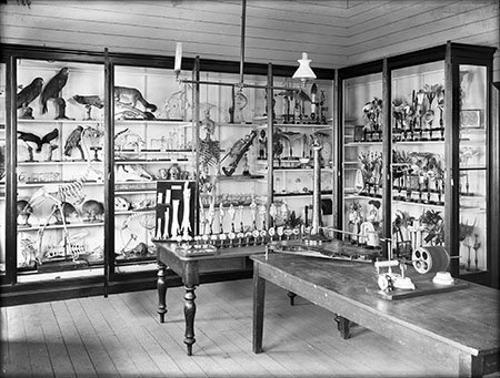
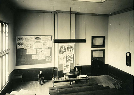
Once acquired, European models and wall charts were employed in lectures and encountered by the colonial public in universities and museums. References in the colonial press and university archives provide insight into how these visual aids were employed in colonial institutions. In 1890, the Natural Sciences Department of Auckland University moved into new premises in Parliament Street, and was featured in the local press.[67] An illustrated article in the New Zealand Graphic depicts Algernon Thomas lecturing in front of a number of wall charts with cases of models to the side.[68]
University of Sydney Archives G3.224.0333
Professors at the University of Sydney also advocated the use of visual aids in their lecture halls and laboratories. T Anderson Stuart, professor of anatomy and physiology at the University of Sydney, recounts,
I certainly developed the use of models to a very great extent. They practically amount to diagrams in three dimensions, and, as they are very often working models, serve to show in a very remarkable or simple way many points in physiology.[69]
On wall charts, he ‘prefer[red] the old method of making diagrams which show the essential part, and which are always before the class, and can be exhibited in full daylight … over lantern slides’, explaining, ‘there are always some fellows in class who will make a disturbance when the room is more or less darkened’. [70]
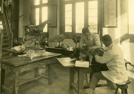
An article on the new biological laboratory erected at Canterbury College in 1896 gives us a further idea on the presentation of models and charts in these new scientific facilities:
The interior of the building is fitted up with all the latest improvements … At the back there are several glass cases containing model specimens in gelatine, papier-mâché and wax, of the vegetable kingdom … They are most ingeniously and delicately constructed, and, being clever reproductions of actual specimens, will be very useful for lecture purposes, both scientific and popular … Leaving the vegetable kingdom, we come to some forms of the animal kingdom. Therein a series of wax models showing the development of the chicken … one showing the development of the heart, and the other that of the ear. Even to those who are not scientists these models must be extremely interesting, on account of the ingenuity shown in their contrivance alone. It is the intention of Professor Dendy to deliver a course of popular lectures, and these models will be of great use for illustrations.[71]
William Haswell similarly supported the use of models. In a handwritten lecture from 1909, entitled, ‘Introductory matter’, he stressed, ‘It is advisable to work as much as possible with the object of study actually before us or failing that, clearly represented to the eye by a diagram or model’.[72] He used images of the Ziegler models he purchased as illustrations in his 1897 A Text-Book of Zoology, co-authored with Thomas Jeffery Parker, professor of zoology at the University of Otago and curator of the Otago Museum.
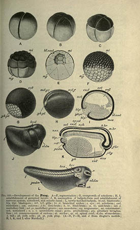
Image from the Biodiversity Heritage Library. Digitised by University of California Libraries
Wall charts also feature in the description of these new biology facilities. At Canterbury College, ‘two large automatic frames, made of slats on broad tapes hang down the wall, on which diagrams can be put. As many as eighteen or twenty illustrations can thus be shown at a time’.[73] In a 1901 letter to W Baldwin Spencer in Melbourne about the new biology laboratory at Sydney University, Haswell discusses the logistical difficulties involved in displaying and storing such charts:
Diagram screens and arrangements for storing the diagrams have been the most troublesome. I am working out a scheme for managing this part of the business on the lines that are followed at Strasburg, but with some modifications. Each of the diagrams that is in constant use for the first year lectures will be provided with a wooden bar bearing hooks by which it can be attached to the screen, and by which it can be hung up in its place in the diagram room.[74]
The mention of popular lectures in some of these excerpts suggests that models and charts were also displayed for the edification of the general public. In 1881 Haswell delivered the 15th of a series of lectures on zoology at the Linnean Society of New South Wales’s rooms in the Garden Palace on the structure, physiology, and life-history of the frog ‘illustrated by a fine series of wax models kindly lent by the trustees of the [Australian] Museum’.[75] At the Royal Society of New South Wales’s conversazione in the Great Hall of the University of Sydney in December 1894 moreover, he showed a ‘series of the stages of chick embryos mounted on a revolving stage’ as well as various plant models.[76] Similar public events were held under Algernon Thomas at a series of ‘At homes’ at Auckland University, where ‘guests wandered ad lib through the laboratories and class rooms’.[77] At a conversazione at the University of Melbourne in 1890, visitors to the biological lab could see ‘a number of coloured diagrams used in illustrating biological lectures’ as well as a collection of German wax models.[78]
Colonial production
European models and wall charts were integral to teaching biology at Australian and New Zealand institutions. However, since the majority of flora and fauna represented in models and charts were not naturally occurring in the colonies, these visual aids represented the triumph of Imperial science over indigenous nature. Imported models and wall charts were supplemented out of necessity by handmade teaching aids devoted to unique Australasian species as well as those geared to colonial economic and agricultural development. In both areas, women practitioners and artists played an active role in producing and promoting local models and charts. The feminine pursuits of flower painting and botanical illustration that were part of a typical middle-class ornamental education provided professional and amateur women artworkers with a relevant and desirable skillset within the professionalising spheres of colonial scientific communities, which lacked the developed networks of specialised practitioners that governed the production of scientific visual aids in Europe.[79] Moreover, women were also active participants in intercolonial and international exhibitions in the late 19th and early 20th centuries, wherein the representation of Australian nature was increasingly equated with that of the nation. Through the display of models and diagrams of native flora and fauna, in addition to a wide variety of women’s work featuring local products, women, as curator Martha Sear has argued, were connected ‘to the processes of developing Australia’s unique resources’ and ‘played a significant role in the production of a national culture’.[80]
Scientific visual aids produced in the colonies were employed in teaching and researching the local natural environment. In the 1890s, as Linden Gillbank has observed, ‘a pro-evolutionary Australasian-based investigation of indigenous biotas was developing’ with ‘economic incentives to investigate the indigenous flora and an evolutionary incentive to examine the native fauna’.[81] At Auckland University, Algernon Thomas discussed the need to catalogue the special Australian flora and fauna while, at the University of Sydney, anatomists James Wilson and JP Hill promoted the study of the anatomy and embryology of native fauna, especially monotremes, and William Haswell researched local marine invertebrates.[82]
It is possible that local Australian species were used to teach anatomy during dissection, although it was the exotic species displayed for teaching. This is suggested in a series of models used at the University of Sydney showing the development of the crayfish. The last of these models is labelled Euastacus serratus, which is a freshwater spiny crayfish found in the rivers of eastern Australia. But this label is incorrect, as the models are clearly based on illustrations published in Thomas Huxley’s The Crayfish: An Introduction to the Study of Zoology (1880), which show the developmental stages of Astacus fluviatilis, the common European crayfish.
To facilitate such research, a number of colonial scientists, or their technicians, made their own diagrams, including John Halliday Scott, professor of anatomy and physiology at Otago University, who was also a practising watercolour artist and painted his own wall charts for anatomical teaching, which continued to be used after his death.[83] The McGregor Museum contains a number of hand-drawn charts from the 1880s, which embody the stylistic conventions of contemporary German charts, presumably made by scientists or illustrators at the University of Auckland, the most predominant of whom used the signature ‘qvp’.[84]
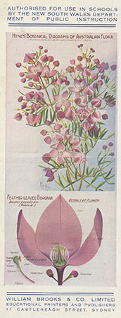
In 1913 Miss Sarah Hynes, a former botanical assistant at the Sydney Botanic Gardens and the Technological Museum, who was appointed a special instructor in science to several schools under the New South Wales Department of Public Instruction, gave a paper at a meeting of the Association for the Advancement of Science held in Melbourne on the benefits of teaching aids depicting local flora:
At the outset it seemed to me only right that in addition to morphology and plant physiology, our native flora should be studied in preference to that of Europe … I was confronted with the difficulty of obtaining wall charts of our native flora to illustrate my lectures. I found there were none procurable. Mrs. Rowan, the distinguished flower painter, came to my assistance, and painted for me an artistically, beautifully, and scientifically accurate series of botanical charts. They have been adopted by the Department of Public Instruction of New South Wales and are being hung in the schools. They are both decorative and instructive and the first of their kind in Australia.[85]
Intended to be published in instalments by William Brooks and Company, Ltd., Sydney, the first set of these charts, measuring 57.15 by 69 centimetres and sold mounted and ready for hanging ‘at the cost of less than a sixpence a week’, comprised 36 studies of native flora, including the honey-flower, native rose, and the feather-leaved boronia, by Ellis Rowan, who had been awarded several medals for her botanical compositions at international exhibitions.[86] ‘Under each plate of the flower proper’, according to an article on the charts in the Evening News, ‘the details are given, and students of botany are enabled to gain the requisite knowledge in the easiest and pleasantest fashion.’[87]
Ellis Rowan Cutting book [manuscript], National Library of Australia MS2203
An illustrated brochure advertising the charts features a number of endorsements from Australian professors and scientists, including praise from Alfred James Ewart, professor of botany and plant physiology at Melbourne University and government botanist of Victoria; Dr JH Johnstone, lecturer in biology, Queensland University; Miss Jean White, lecturer in the Department of Botany, Melbourne University; and JA Turner, Director of Technical Education in New South Wales. These endorsements stress the usefulness of the wall charts in teaching botany and educating students in the plant side of nature study, their role in inculcating a love of local nature, and their aesthetic merit as ‘unique examples of Artistic Flower Painting’.[88] While Hynes’s charts were principally designed for use in school education, diagrammatic series of local flora were also employed at the university level, as revealed in an early 20th-century photograph of a lecture hall at Sydney University featuring a chart of gum leaves and gum leaf blossoms.
University of Sydney Archives G3.224.1006.5
In addition to advocating the production of wall charts of native flora, Hynes also advocated the use of models, highlighting the lack of local series of Australian flowers and fruit:
Another difficulty which I encountered was the absence of models for teaching purposes of our native flowers and fruits. When technical assistant of the Technological Museum, there were models made under my direction of some common flowers, but the material (plaster of Paris) was unsuitable, and very few were made. I am now negotiating for a series of models to be made in suitable material.[89]
It was particularly in the area of economic botany that locally made models were required for teaching at technical colleges or for public instruction in museums. Unusually, the Museum of Economic Botany in the Adelaide Botanic Gardens, under director Dr Richard Schomburgk, acquired a ‘pomological cabinet’ of 360 papier-mâché models, mainly apples and pears, made by German company Heinrich Arnoldi & Co. over the years 1856 to 1899.[90] These models, accurately named by reputable pomologists, were seen as important references to ensure correct naming of species or possible imports.
This German collection was supplemented by locally produced wax models of fruit, a number of which were made by Adelaide artist Miss Ellen Lucy Gray and displayed at the Philadelphia Exhibition and the Paris Exposition in 1878.[91] Gray’s contributions can be seen as an extension of the feminine amateur tradition of modelling botanical specimens in wax, which were often submitted to horticultural society displays and international exhibitions. The natural products of New South Wales on show at Paris Universal Exhibition 1867, for instance, included wax models of Australian flowers by Elizabeth Podmore, Sydney, and wax bush flowers of Australia by Mrs Johns.[92]
In Victoria, the Industrial and Technical Museum had amassed by 1878 a collection of wax fruit used as reference copies against which ‘every fruit that can establish its claim to novelty or exceptional excellence’ or the claims of ‘new pretenders’ could be tested.[93] Two models were made of each piece of fruit, one model for the Department of Agriculture and one for the museum, which was placed on display for the education of the public, with Thomas McMillan working as the principal modeller at the museum in the late 19th century.[94] A number of these models of Victorian fruit were exhibited at the London, Philadelphia, Paris and Calcutta international exhibitions, as well as in the intercolonial exhibitions in Melbourne and Sydney. Two albums survive in the museum’s collection of hand-painted illustrations of fruit models: one of examples produced between 1872 and 1879, and another that contains hand-painted references of models of apples, apricots, peaches, pears and plums, compiled by Miss McMillan in 1904.[95]
Miss McMillan became a professional modeller in her own right and, as Mrs AG Goodman, was employed at the Technological Museum in Sydney by Maiden, who had seen the models in Melbourne. By 1885 she had modelled more than 300 fruits and vegetables, as well as plaster meteorites and gold nuggets.[96] Located then in Agriculture Hall, the Outer Domain, the museum displayed a collection of greatly enlarged models of flowers and wax models of fruits grown in this colony.[97] These models and the collection in Melbourne ‘were very frequently referred to, and inspected by those engaged in the fruit industry’, prompting the Tasmanian Museum to address ‘the need of a collection of wax models, correctly coloured, of Tasmanian fruits’ of products grown for market and export, a collection to be ‘both educative and practically useful’.[98] It was reported that the Victorian artist was away for a year or two, but the Sydney Technological Museum had an artist who could do the work, at a cost of 8 shillings per specimen.
In addition to featuring in museum collections, models of flowers were also produced at the Sydney Technical College, the biology course of which was mainly for teacher training (primary and secondary), but also practical gardeners and nurserymen and agriculture students.[99] Modeller and teacher Arthur Edward Rice and colourist Charles Tom produced a collection of botanical models for the college. Rice, a former student, had become a teacher in 1904, before joining the Wunderlich manufacturing company as a modeller, while Tom taught at the college from 1890 to 1916.[100] Such local models and charts were limited to their own area; illustrative, they were deemed generally accurate but remained distinct from scientific models produced for academic study as their makers did not work closely with scientists.
Conclusion
The emergence of biology as a professional scientific discipline in the late 19th century was accompanied by a change in its teaching. Comparative morphology and embryology, based on microscopical observation and experiment, were taught with practical training in the laboratory as the place of learning. Visual aids, in particular models and wall charts, became a vital part of this teaching, by helping students understand what they were seeing. This approach appeared first in Germany, with England lagging, until Thomas Huxley at South Kensington in the 1870s introduced laboratory-based teacher training.
In Australian and New Zealand, the new university departments of biology imported teachers trained in this new way of teaching, who brought European scientific teaching aids into their classrooms. The need for models and charts to teach economic botany based on native flora and fauna saw a small number of locally produced products, but never on the scale or with the scientific authority of the European trade. A distinguishing trait in the colonial production of scientific teaching aids, however, was the prominent role of women practitioners in manufacturing a national image of Australia through its natural resources.
After the Second World War, the teaching of biology changed dramatically. Fuelled by ‘the shift from the study of life grounded in the natural history tradition to modern experimental biology’, the production and use of models, charts and specimen sets declined from the late 1950s with existing university and museum collections put into storage or discarded.[101] With the development of more dynamic exhibitions spaces in mid-century museological practice, older cases were retired and collections of teaching aids were increasingly removed from public view.
In the late 20th and early 21st centuries, however, this scientific visual culture has reappeared, particularly in the sphere of contemporary art. Embracing the aura of the Wunderkammer and the return to craft, contemporary artists such as the Sydney-based, Columbian-born María Fernanda Cardoso, have resurrected the practice of making models inspired by nature while drawing upon evolving image technologies.[102] The legacy of biological visual aids has a palpable presence in this work, suggesting that current artistic practice continues to react to the marvel of visualising nature in material form.
In tandem with such contemporary model making, collections of historic models and charts are being rediscovered and brought back into the light. Often they have to be re-identified as they have been transferred to the museum store from the laboratory. Research into this instrumental genre of late 19th- and early 20th-century scientific educational visual culture is informing a new understanding of its production and dispersal. As categorically evasive objects that subscribe to the history of science, the history of education, museology, art history and material culture studies, models and wall charts require an interdisciplinary and collaborative approach. This approach, moreover, must be sensitive to the shifting significance of such visual aids over time, from their original employment as cutting-edge teaching devices, many reflecting the latest research in biology in the late 19th- and early 20th-centuries, to scientifically and technologically obsolete educational artefacts that have recently acquired new aesthetic significance.
This paper has been independently peer-reviewed.
Endnotes
1The research for this paper began with an exhibition curated by Jan Brazier at the Macleay Museum, the University of Sydney, in 2013, True to Form: Models for Teaching Science, which explored models used for teaching at the university. A joint paper was presented by Brazier and Molly Duggins, ‘Visualising nature in the classroom: German scientific models and wall charts in Australia and New Zealand’, at the AAANZ conference, Melbourne, 2013.
Lynn Nyhart, Modern Nature: The Rise of the Biological Perspective in Germany, University of Chicago Press, London, 2009, p. 17. See also Lynn Nyhart, ‘Natural history and the “new” biology’, in N Jardine, JA Secord & EC Spary (eds), Cultures of Natural History, Cambridge University Press, Cambridge, 1996, pp. 426–43.
2 Anne Secord, ‘Botany on a plate: Pleasure and the power of pictures in promoting early nineteenth-century scientific knowledge’, Isis, vol. 93, no.1, March 2002, 28–57 (p. 52).
3 Tony Bennett, ‘Speaking to the eyes: Museums, legibility and the social order’, in Sharon Macdonald (ed.), The Politics of Display: Museums, Science, Culture,Routledge, New York, 1998, pp. 25–35. The South Kensington Museum was predecessor to the Science Museum.
4 Brian Foster, Jack Rattenbury & John Marbrook, A History of Biology at Auckland University 1883–1983, Research report, Department of Biology, University of Auckland, 1983, p. 17.
5 Geoffrey N Swinney, ‘Enchanted invertebrates: Blaschka models and other simulacra in National Museums Scotland’, Historical Biology, vol. 20, no. 1, 2008, 39–50 (p. 40).
6 Nyhart, Modern Nature, p. 27.
7 Nick Hopwood, Embryos in Wax: Models from the Ziegler Studio, Whipple Museum of the History of Science and the Institute of the History of Medicine, Cambridge and Bern, 2002, pp. 34–5; Massimiano Bucchi, ‘Images of science in the classroom: Wallcharts and science education 1850–1920’, British Journal for the History of Science, vol. 31, 1998, 161–84 (p. 165).
8 Hopwood, Embryos in Wax, p. 35. Hopwood adds, ‘Magnification brought together very different objects at a size suitable for demonstration, and – as the products of a single firm – in similarly standardized forms’.
9 Secord, ‘Botany on a plate’ (p. 32)
10 Hopwood, Embryos in Wax, p. 36.
11 Swinney, ‘Enchanted invertebrates’ (p. 41); Nick Hopwood, ‘Plastic publishing in embryology’, in Soraya de Chadarevian & Nick Hopwood (eds), Models: The Third Dimension of Science,Stanford University Press, California, 2004, p. 178 .
12 Ann B Shteir, “‘Fac-similes of nature”: Victorian wax flower modelling’, Victorian Literature and Culture, vol. 35, No. 2, 2007, 649–61 (p. 660); Swinney, ‘Enchanted invertebrates’ (p. 41).
13 Lorraine Daston, ‘The Glass Flowers’ in Lorraine Daston (ed.), Things that Talk: Object Lessons from Art and Science , Zone books, New York, 2004, p. 225: ‘They are at once undeniably artificial and flawlessly natural, in the tradition of mimesis that extends back to Pliny’s story.’
14 Celeste Olalquiaga, The Artificial Kingdom: A Treasury of the Kitsch Experience, Bloomsbury, New York, 1998; Daston, ‘The Glass Flowers’, p. 224.
15 Swinney, ‘Enchanted invertebrates’ (p. 42).
16 E Ray Lancaster, ‘Dealers in zoological specimens and models’, Nature, vol. 16, no. 416, 18 October 1877, 521. Lancaster was later director of the Natural History Museum from 1898 to 1906.
17 For an overview of the main model makers, see Sabine Hackethal, ‘The Blaschka models of the Humboldt University of Berlin and their historical context’, Historical Biology , vol. 20, no. 1, March 2008, 19–28; in particular, the makers the Blaschkas, Zieglers, Weisker, Loth, Osterloh and Sommer .
18 Robert Bud, ‘Responding to stories: The 1876 Loan Collection of Scientific Apparatus and the Science Museum’, Science Museum Group Journal, issue 1, Spring 2014, http://doi.crossref.org/10.15180/140104.
19 Hopwood, ‘Plastic publishing’, p. 185.
20 Hopwood, Embryos in Wax, p. 54.
21 Catalogue of the Special Loan Collection of Scientific Apparatus at the South Kensington Museum,London, 1877, 3rd edition, item 3839, p. 984.
22 Hopwood, Embyros in Wax, pp. 31, 191; Hackethal, ‘The Blaschka models’ (p. 22).
23 Otago Daily Times 8 February 1882, p. 1; ‘Otago Institute’, 20 September 1893, p. 3. See Hopwood, Embryos in Wax, pp. 119–56,for reproduction of a Ziegler catalogue.
24 Press, (Christchurch), 13 March 1896, p. 3.
25 Bucchi, ‘Images of science’ (pp. 169, 171); Daston, ‘The Glass Flowers’, p. 233.
26 Daston, ‘The Glass Flowers’, p. 233.
27 The 1878 catalogue is viewable online; HA Ward’s Natural Science Establishment, Catalogue of Glass Models of Invertebrate Animals,Rochester, New York, 1878, Hathi Trust Digital Library.
28 Press, (Christchurch), 1 November 1883, p. 6.
29 ‘Some clever glass-blowing’, Hawke’s Bay Herald, 14 December 1883, p. 4.
30 Brisbane Courier, 8 September 1885, p. 3.
31 Catalogue of the Special Loan Collection, item 3864, p. 988.
32 ibid.
33 Dawn Sanders, ‘The death and life of the plant specimen’, in Peter Heering & Roland Wittje (eds), Learning by Design: Experiments and Instruments in the History of Science Teaching, Franz Steiner Verlag, Stuttgart, 2011, p. 161; Graziana Fiorini, Luana Maekawa & Peter Stiberc, ‘Save the plants: Conservation of Brendel anatomical botany models’, The Book and Paper Group Annual , vol. 27, 2008, http://cool.conservation-us.org/coolaic/sg/bpg/annual/v27/bp27-07.pdf, accessed 22 February 2015.
34 Henri Reiling, ‘Über Blaschkas Glasmodelle und die zeitgenössische Naturgeschichte, mit einem Anhang über Brendels botanische Modelle. [On the Blaschkas’ glass models and contemporary natural history, with an appendix on the Brendels’ botanical models.]’, in Michael Kaasch & Joachim Kaasch (eds), Verhandlungen zur Geschichte und Theorie der Biologie, vol. 14, Verlag für Wissenschaft und Bildung, Berlin, 2009, Appendix: English translation online http://members.ziggo.nl/here/blaschka-brendel.html, accessed 17 January 2015.
35 See http://mcgregor.sbs.auckland.ac.nz/2010/12/15/brendel-botanical-models, accessed 19 January 2013. For Otago Museum, see Otago Witness, 10 August 1893, p. 15.
36 Margaret Maria Cocks, ‘Dr Louis Auzoux and his collection of papier-mâché flowers, fruits and seeds, Journal of the History of Collections, vol. 26, no. 2, 2014, 229–48. This article does not deal with human anatomical teaching aids, to which a substantial amount of literature has already been devoted, but on this trade see Anna Maeker, ‘Anatomizing the trade: Designing and marketing anatomical models as medical technologies, ca. 1700–1900’, Technology and Culture, vol. 54, no. 3, July 2013, 531–62.
37 The Technological Museum was the predecessor of the Museum of Applied Arts and Sciences, the Powerhouse Museum. The models, registration number 2796, can be seen at the Powerhouse Museum’s collections online: www.powerhousemuseum.com/collection/database/menu.php. The catalogue and price list for the Auzoux fungi models is item 2005/213/1.
38 Susie Shears, ‘The physick gardener’, University of Melbourne Collections, issue 6, June 2010, 8–13 (p.11).
39 Evening News,12 March 1901, p. 7.
40 Henry T Tribe, ‘Sowerby’s models and “Sowerby Inspiration Models”’ in Bulletin of the British Mycological Society, 18, 1984, 61–4.
41 Tony Kanellos, Imitation of Life: A Visual Catalogue of the 19th Century Fruit Models in the Santos Museum of Economic Botany in the Adelaide Botanic Garden, Wakefield Press, Adelaide, 2013, n.p.
42 Sydney Morning Herald, 26 January 1910, p. 7.
43 Rudolf Schmid, ‘Wall charts (Wandtafeln): Remembrance of things past’, Taxon , vol. 39, no. 3, Aug 1990, 471–2 (p. 471).
44 Bucchi, ‘Images of science’ (p. 184).
45 ibid. (p. 182).
46 Willam Blaxland Benham, Catalogue of Diagrams in the Biological Department of the Otago University Museum, 1902-22, ourheritage.ac.nz | OUR Heritage, accessed February 22, 2015, http://otago.ourheritage.ac.nz/items/show/7811.
47 Bucchi, ‘Images of science’ (p. 172).
48 ibid., p. 182.
49 Biology requisitions book, entry 25 April 1902, G50/7, University of Sydney Archives. The book also contains a reference to Kny charts.
50 The McGregor Museum and the University of Melbourne also have a series of wall charts, drawn in a manner similar to Leuckart’s, by the geologist and palaeontologist Karl Alfred von Zittel (1839–1904), a professor of geology at the University of Munich and the director of the natural history museum.
51 Alison Abbott, ‘Science in culture’, Nature, vol. 421, no. 580, 6 February 2003, 580.
52 Thomas AP Van Leeuwen, ‘Mezzanine art, or the stor(e)y between science and art’, Historical Biology, vol. 20, no. 1, 2008, 63–75 (p. 65).
53 Linden Gillbank, ‘The life sciences: Collections to conservation’, in Roy MacLeod (ed.), The Commonwealth of Science, Oxford University Press, Melbourne, 1988, p. 102; on the foundation appointments, see also Ian Inkster & Jan Todd, ‘Support for the scientific enterprise,1850–1900’, in RW Home (ed.), Australian Science in the Making, Cambridge University Press, Cambridge, 1988, p. 116.
54 Purchases of apparatus and equipment are referred to in the Minutes of the Science Faculty, for example, p. 2; University of Sydney Archives G3/3/1.
55 Foster, Rattenbury & Marbrook, A History of Biology, p. 19.
56 On Friĉ, see Henri Reiling & Tat’jána Spunarová, ‘Václav Friĉ (1839–1916) and his influence on collecting natural history’, Journal of the History of Collections, vol. 17, no. 1, 2005, 25–43. On the purchase by the Australian Museum, see Correspondence, AMS 7, C.1.79.1 and C.10.81.5, Australian Museum Archives.
57 Australian Museum, Annual Report, 1877, p. 2.
58 V Friĉ to A. Liversidge, 1 February 1879, AMS 7, C.10.81.5, p. 27, Australian Museum Archives.
59 AMS 7, C.10.81.5, p. 14. The arrival of the first consignment of 10 cases was recorded by the Sydney Morning Herald,18 July 1881, p. 3. An atlas and diagrams were listed in the proposed items but no other details recorded.
60 Correspondence in the outward exchange letterbooks, AMS 59/1, 1884, pp. 388, 393, Australian Museum Archives.
61 Sydney Morning Herald,17 November 1885, p. 6.
62 ‘The Australian Museum’, Australian Town and Country Journal, 14 August 1897, p. 22; Patricia Egan, ‘A delicate record: Blaschka glass models’ in Feathers of the Gods and Other Stories from the Australian Museum, Australian Museum, Sydney, 2013, pp. 60–1.
63 Correspondence letterbooks, vol. 2, MRS4, pp. 285–6, Powerhouse Museum Archives.
64 ibid., p. 424.
65 Letter from Rabone Feez & Co., 10 April 1884, reporting from their Hamburg correspondent, ibid., p. 479.
66 C. Vetter to G. Masters, 30 Nov 1890, Macleay Museum Archives.
67 Foster, Rattenbury & Marbrook, A History of Biology, pp. 18-19.
68 ‘Auckland University, the college by the sea: A short study by a sometime student – in two parts’, New Zealand Graphic, 13 October 1894, 340–1. The article also notes that Thomas made illustrative drawings on the blackboard ‘of quite unusual excellence’ (p. 340). For more on blackboard illustrations, see Caitlin Donahue Wylie, ‘Teaching nature study on the blackboard in late nineteenth- and early twentieth-century England’, Archives of Natural History, vol. 39, no. 1, 2012, 59–76.
69 William Epps, Anderson Stuart, M.D. , Angus & Robertson, Sydney, 1922, p. 4.
70 ibid., p. 5.
71 ‘New Biological Laboratory’, Press, 13 March 1896, p. 3.
72 ‘Introductory Matter’, 2nd lecture 1909, attrib. Haswell, P168/1, series 4, University of Sydney Archives.
73 ‘New Biological Laboratory’, p. 3
74 Haswell to Baldwin Spencer, 12 August 1901, MSS29, p. 106, Sir Walter Baldwin Spencer Papers, Mitchell Library.
75 Sydney Morning Herald, 25 November 1881, p. 8.
76 ‘Conversazione’, Journal and Proceedings of the Royal Society of New South Wales, vol. 28, 1894, 334–9, (p. 336). Various plant models and zoological diagrams were also exhibited.
77 Foster, Rattenbury & Marbrook, A History of Biology, p. 19.
78 Argus, 14 January 1890, p. 7.
79 Caroline Jordan, Picturesque Pursuits: Colonial Women Artists and the Amateur Tradition, Melbourne University Press, Melbourne, 2005, p. 184.
80 Martha Sear, ‘“Common neutral ground”: Feminising the public sphere at two nineteenth-century Australian exhibitions of women’s work’, in Kate Darian-Smith, Richard Gillespie, Caroline Jordan & Elizabeth Willis (eds), Seize the Day: Exhibitions, Australia and the World, Monash University ePress, Clayton, Vic, 2008, pp. 470, 475.
81 Gillbank, ‘The life sciences’, pp. 102, 104.
82 Foster, Rattenbury & Marbrook, A History of Biology, p. 17; Patricia Morison, ‘Wilson, James Thomas (1861–1945)’, Australian Dictionary of Biography, National Centre of Biography, Australian National University, http://adb.anu.edu.au/biography/wilson-james-thomas-9140, accessed 7 March 2015.
83 See www.teara.govt.nz/en/biographies/2s7/scott-john-halliday,accessed 22 January 2015.
84 See http://mcgregor.sbs.auckland.ac.nz/category/historic-charts/zoology-charts/page/5/, accessed 22 January 2015.
85 ‘Nature study: Teaching of botany: An interesting paper’, Evening News, 22 January 1913, p. 6.
86 William Brooks & Co. Ltd, ‘Hynes’ botanical diagrams of Australian flora’ (brochure), 1913, Ellis Rowan cutting book [manuscript]. MS2203, National Library of Australia; Caroline Jordan, ‘Tom Roberts, Ellis Rowan and the struggle for Australian art at the Great Exhibitions’, in Seize the Day: Exhibitions, Australia and the World, pp. 496–527.
87 ‘Australian flora’, Evening News, 4 August 1911, p. 8.
88 William Brooks & Co. Ltd, ‘Hynes’ botanical diagrams’.
89 Evening News, 22 January 1913, p. 6.
90 Kanellos, Imitation of Life, p. 9.
91 Obituary of Mrs RS Thompson (Ellen Lucy Gray), South Australian Register, 8 July 1878, p. 5.
92 New South Wales Exhibition Commissioners, Catalogue of the Natural and Industrial Products of New South Wales Forwarded to the Paris University Exhibition of1867, Government Printer, Sydney, 1867, p. 28.
93 ‘The sketcher’, The Australasian, 23 February 1878, p. 232.
94 See http://museumvictoria.com.au/collections/themes/1187/wax-fruit-collection, accessed 23 January 2015.
95 http://museumvictoria.com.au/collections/items/403798/album-wax-fruit-models-miss-mcmillan-1904. Fruit and vegetable models were produced by the museum until about 1960, when replaced by colour photography.
96 See www.powerhousemuseum.com/insidethecollection/2010/01/calling-all-apple-experts, accessed 23 January 2015.
97 Sydney Mail and New South Wales Advertiser,12 June 1886, p. 1212.
98 ‘Tasmanian fruits’, Mercury, 27 March 1909, p. 11.
99 A Quarter Century of Technical Education in New South Wales, Government Printer, Sydney, 1909, p. 174.
100 See online collection catalogue for the Powerhouse Museum where the models are now held.
101 John L Rudolph, ‘Teaching materials and the fate of dynamic biology in American classrooms after Sputnik’, Technology and Culture, vol. 53, no. 1, January 2012, 1–36 (p. 18).
102 Focusing on the microscopic world of insect genitalia, Cardoso has created a number of artworks based on traditional modelling processes combined with rapid 3D prototyping technology, which were exhibited in her The Museum of Copulatory Organs (MoCo) at the 2012 Sydney Biennale.
 ISSN 1833-4946